Cheek microscope animal rsscience lesson Draw the diagram of cheek cells and label the parts. Cell cheek observation stained nucleaus safranin
SBI3U
Diagram of composite cell Cheek cells Diagram of human cheek cell and onion cell
Draw the diagram of cheek cells and label the parts.
Drawing cheek cell labelled human biological parts followed rules basic must there some when exportedCell onion cheek human diagram diagrams Cheek cellsBbc bitesize.
Which of the following components are seen in slide of human cheek cellCells cheek bbc science revision bitesize ks3 systems Observation of nucleaus in cheek cellPolymath at large: the little things that keep us going.

Cheek cells human keep going things little epithelial mitochondria polymath large
Cells cheek microscope human under cell animal membrane epitheliumCells cheek microscope blue cell epithelial methylene ks3 stained structure bbc revision bitesize biology ultrastructure drawing observing magnification Cell structures & functionCell cheek single composite diagram anatomy human membrane guws medical.
Cheek cell human temporary stained cells mounts prepare epithelial lab results layer work discussionEal biology: different types of cells Cheek magnification typical nucleus photographedCells cheek biology eal liver.
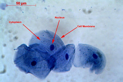
Cheek cells
Diagram of. cheek cellCheek cell cells human membrane animal plant lab eukaryotic epithelium squamous post ppt powerpoint presentation obvious nuclei cytoplasm Flashcards table on bio lab midtermCheek cell human draw labelling correct.
My cheek cellsCheek cell human label parts brainly following answer Label the following parts of human cheek cellCell cheek cells observed wall components slide seen human following under which when yes they.

Cheek correct labelling brainliest ppz
Cell cheek animal structuresSolved using this table from the size estimation module, Lesson 2: mount a slide & “look at your cheek cells“Cheek sel hewan organel fungsi dimiliki.
Sbi3uDiagram of. cheek cell Cheek biologycornerLab cheek cells epithelial human nucleolus cytoplasm midterm bio flashcards membrane plasma nucleus labs.

Draw the human cheek cell with correct labelling
Cell structureCells cheek cell bubble air blue stain To prepare stained temporary mounts of human cheek cellDraw the human cheek cell with correct labelling.
Cheek cell bacteria cells human nucleus membrane using single bacterial been solved determine prokaryoticCheek diagram Human cheek cells under the microscopeCells cheek cell labeled 100x 2009 400x magnified.

SBI3U

cell-cheek-03

Lesson 2: Mount a Slide & “Look at Your Cheek Cells“ - Rs' Science

Solved Using this table from the Size Estimation module, | Chegg.com

BBC - KS3 Bitesize Science - Cells to systems : Revision, Page 2

Cell Structures & Function - AG.& ENVIRONMENTAL SCIENCES ACADEMY

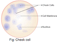
Cheek Cells
